Substrate Control of Dendrite Growth
As animals grow, dendrite arbors of many neurons must expand proportionally to sustain proper connectivity and maintain coverage of their receptive field. Despite the prevalence of this phenomenon, particularly in sensory systems, how dendrite arbor expansion is coordinated with target receptive field expansion is largely unknown. Using genetic screens for mutations that uncouple growth of dendrites and their substrate we have identified a series of substrate-derived cues that coordinate dendrite-substrate growth, in part by restricting dendritic structural plasticity

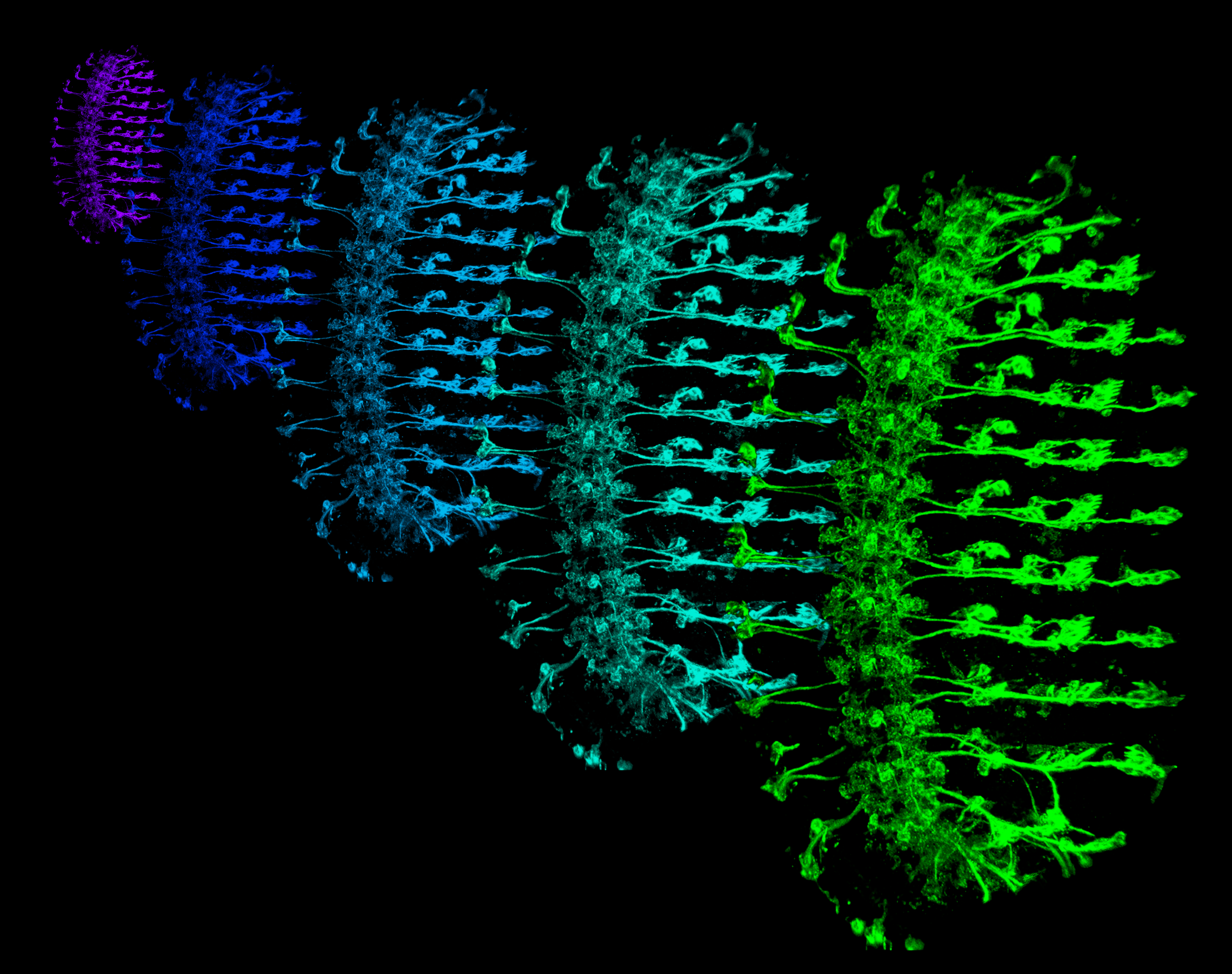
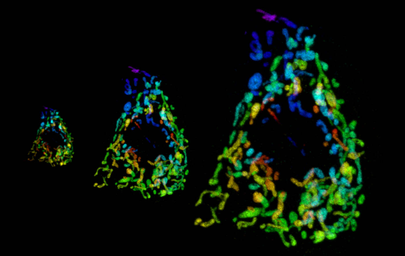
Scalar expansion of dendrite arbors. As animals grow, expansion of dendrite arbors and their substrate must be coordinated to maintain receptive field coverage. (A) Goldfish ganglion cells maintain constant coverage of the growing retina. Dendritic territories (shaded polygons) and cell bodies (black oval) are depicted for Type 1.2 ganglion cells from two fish, one 3.9 cm in length, the other 14 cm in length. Adapted from (Hitchcock, 1987). (B) Drosophila somatosensory neurons establish dendritic coverage of the body wall by Day 2 of development and maintain this coverage even as larvae grow more than 10-fold in linear dimensions. Confocal images show dendrite arbors on Day 1, 2 and 4 of development; dendritic territories of individual neurons are shaded.
Dendrite-epidermis interactions
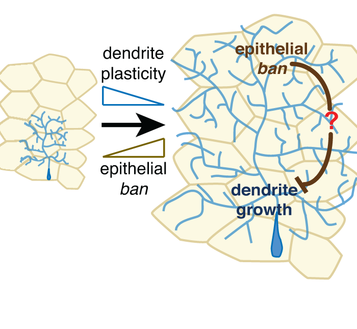
We have found that physical coupling of epithelial cells and dendrites in the form of dendrite invagination into epithelial cells progressively increases during development, directly coupling dendrite growth to substrate expansion and constraining structural plasticity of dendrites. Notably, similar dendrite-epidermis interactions have been described in diverse sensory systems (worm, fly, fish), suggesting that these mechanisms are likely functional in higher organisms as well.

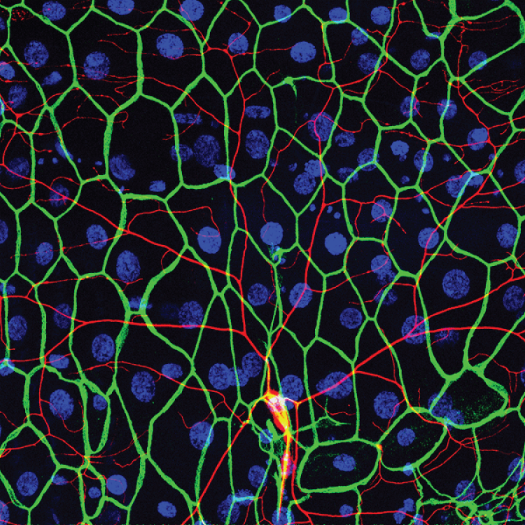
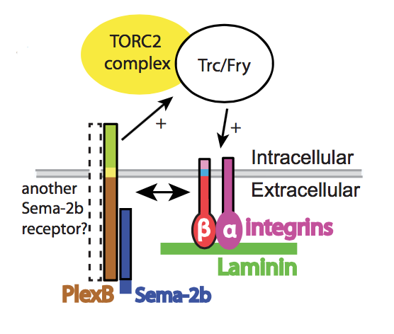
Drosophila larval sensory dendrites (green) grow over a monolayer of epithelial cells (gray) in a mostly two-dimensional plane, attached to a collagen-rich basement membrane (BM, tan) along the basal surface of epithelial cells by virtue of neuronal Integrin (magenta) and Ret (blue). The basement membrane is likewise tethered to the epidermis by epithelial Integrins. The majority of dendrites are positioned at the basal plasma membrane of epithelial cells (BM-attached dendrites; shown at high magnification in a TEM micrograph at right), but a small portion of dendrites become embedded in the epidermis.